NanoACi & Mytocel MSK

Two treatments.One important difference.

Mytocel MSK prepares a patient’s own auricular cartilage as autologous micrografts. NanoACi takes that cellular step and adds a cell-free ChondroFiller scaffold and PRF in a one-stage, needle-delivered technique developed by Professor Lee. See the process, the distinctions and the right next step for you.

Point-of-care preparationOne planned sittingNeedle-deliveredNo laboratory cell cultureNo arthroscopy for NanoACi
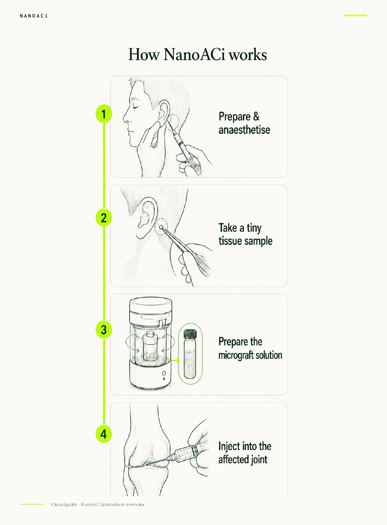
Four-step overview of the NanoACi point-of-care procedure

Evidence for an individual component does not automatically become evidence for the complete protocol.

The evidence layers are reported separately below.

Start with the distinction

Mytocel MSK supplies the micrografts. NanoACi is the wider technique.

The fastest way to understand the page is to keep the cellular treatment separate from the three-part protocol that uses it.

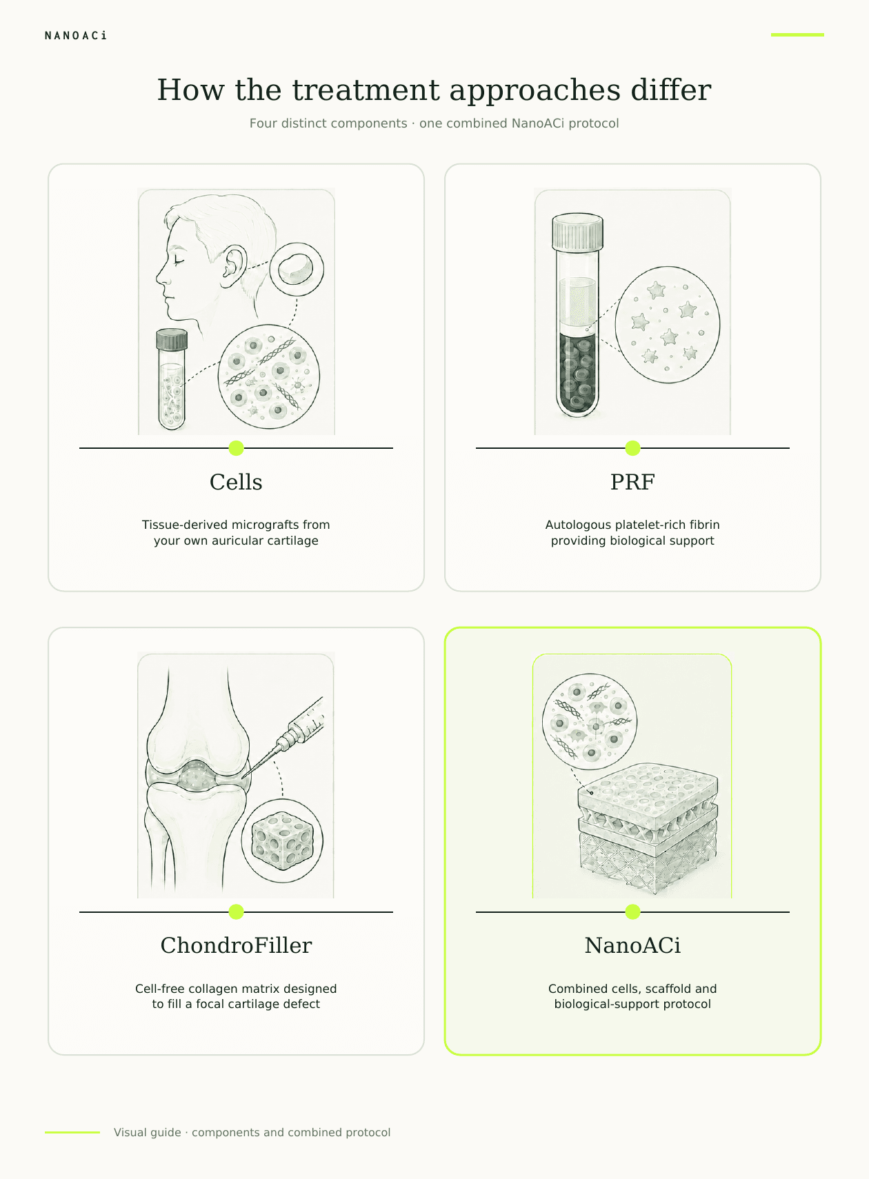
Visual comparison of the cellular, scaffold and PRF roles within NanoACi

Professor Lee technique

NanoACi

Non-Arthroscopic Needle-Delivered One-Stage Autologous Chondrogenic Injection

A single-session injection technique developed by Professor Lee. It uses autologous cartilage micrografts, PRF prepared from the patient’s blood and a ChondroFiller collagen scaffold.

NanoACi is the name of the clinical technique. It is not the name of a single product or device.

Explore the dedicated NanoACi site
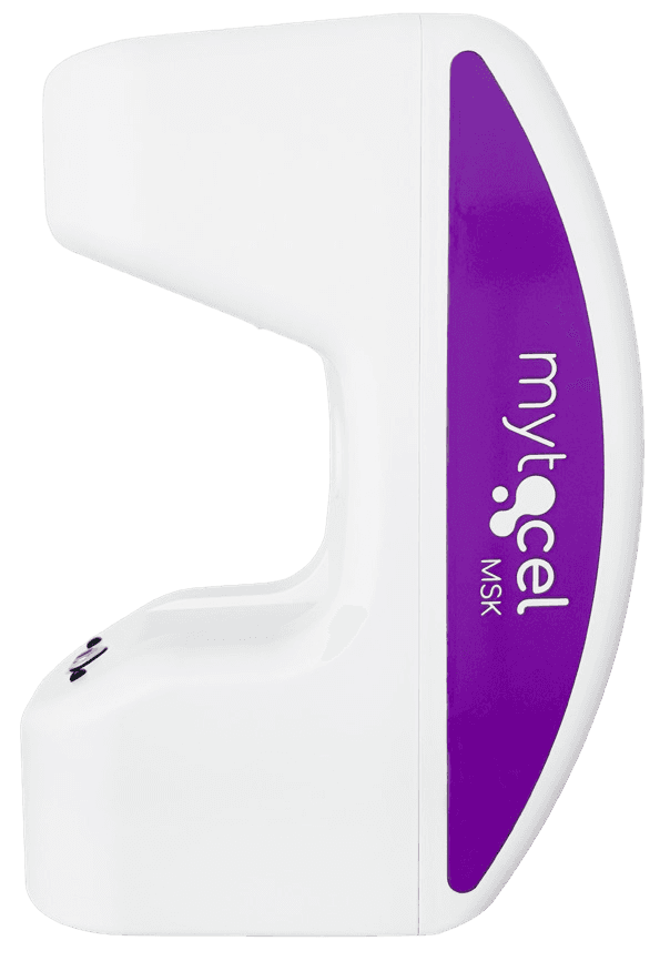
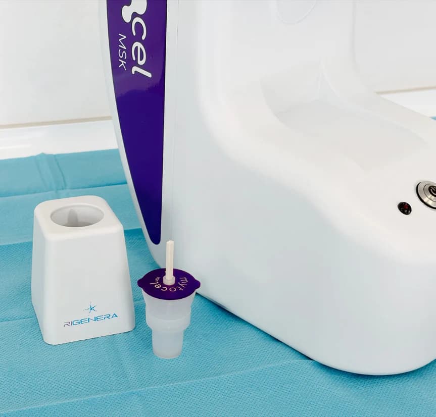
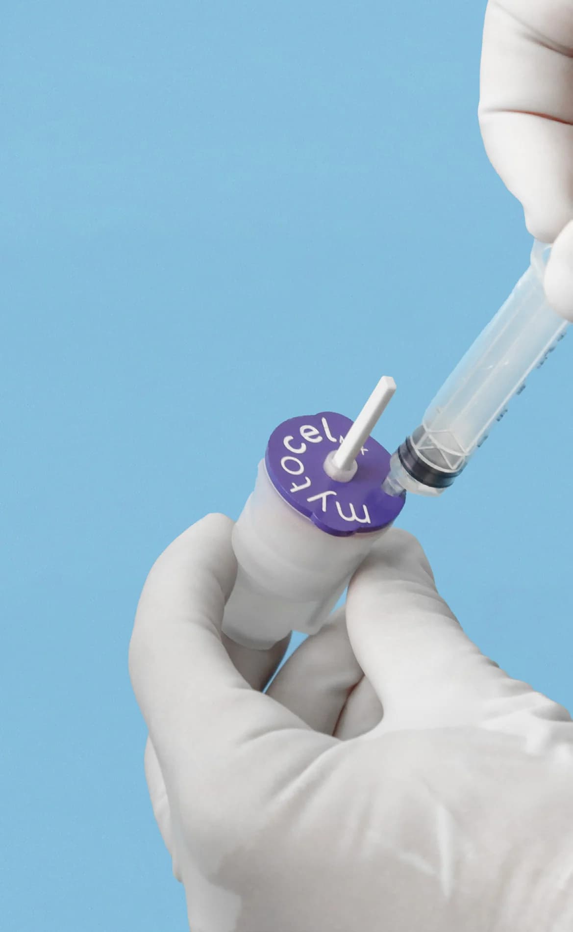
Mytocel MSK point-of-care processing device

Autologous micrograft treatment

Mytocel MSK

A single-session treatment in which tiny samples of the patient’s auricular cartilage are mechanically prepared at point of care into autologous micrografts, without enzyme digestion or laboratory cell culture.

Mytocel MSK can be considered as a standalone treatment and supplies the cellular micrograft step within NanoACi. The underlying Regenera Activa technology is manufactured by Remedi.

Read the patient treatment guide

Side-by-side

What changes when Mytocel MSK becomes part of NanoACi?

Mytocel MSK is a complete treatment in its own right. Inside NanoACi, it becomes one biological step within a wider, surgeon-led delivery sequence.

What it is

Mytocel MSK

An autologous cartilage-micrograft treatment.

NanoACi

Professor Lee’s wider needle-delivered cartilage technique.

Patient-derived input

Mytocel MSK

Tiny auricular-cartilage samples, mechanically prepared at point of care.

NanoACi

The same cartilage-micrograft step, plus PRF prepared from the patient’s blood.

Scaffold

Mytocel MSK

No ChondroFiller scaffold forms part of standalone Mytocel MSK.

NanoACi

A cell-free Type I collagen ChondroFiller scaffold is a defined component.

Clinical distinction

Mytocel MSK

Can be considered as a standalone point-of-care micrograft treatment.

NanoACi

Sequences cellular material, scaffold and PRF as one non-arthroscopic, surgeon-led protocol.

Evidence position

Mytocel MSK

Published studies examine the underlying autologous micrograft technology.

NanoACi

Its components have relevant evidence; the exact three-part protocol is newer and is being followed through NanoACi 100.

Mytocel MSK · the technique

From a tiny ear-cartilage sample to injectable micrografts

The defining feature is not simply that the material is autologous. It is tissue-specific auricular cartilage, mechanically prepared at point of care and delivered without laboratory culture.

  1. 01

    Prepare and anaesthetise

    The auricular donor area is prepared under sterile conditions and numbed with local anaesthetic.

  2. 02

    Take tiny cartilage samples

    Three small samples are taken from the cartilage of the outer ear. This supplies tissue-specific autologous starting material.

  3. 03

    Prepare the micrografts

    The samples are mechanically disaggregated in the Rigeneracons RS using the N4SA system. There is no laboratory culture, enzyme digestion or multi-week expansion.

  4. 04

    Deliver in the same sitting

    The prepared autologous micrograft suspension is collected and delivered to the target joint as part of the same point-of-care pathway.

Mytocel MSK N4SA system and Rigeneracons point-of-care setup
N4SA processing system with the sterile Rigeneracons device.
Sterile preparation of the Mytocel MSK Rigeneracons device
Mechanical preparation at point of care.
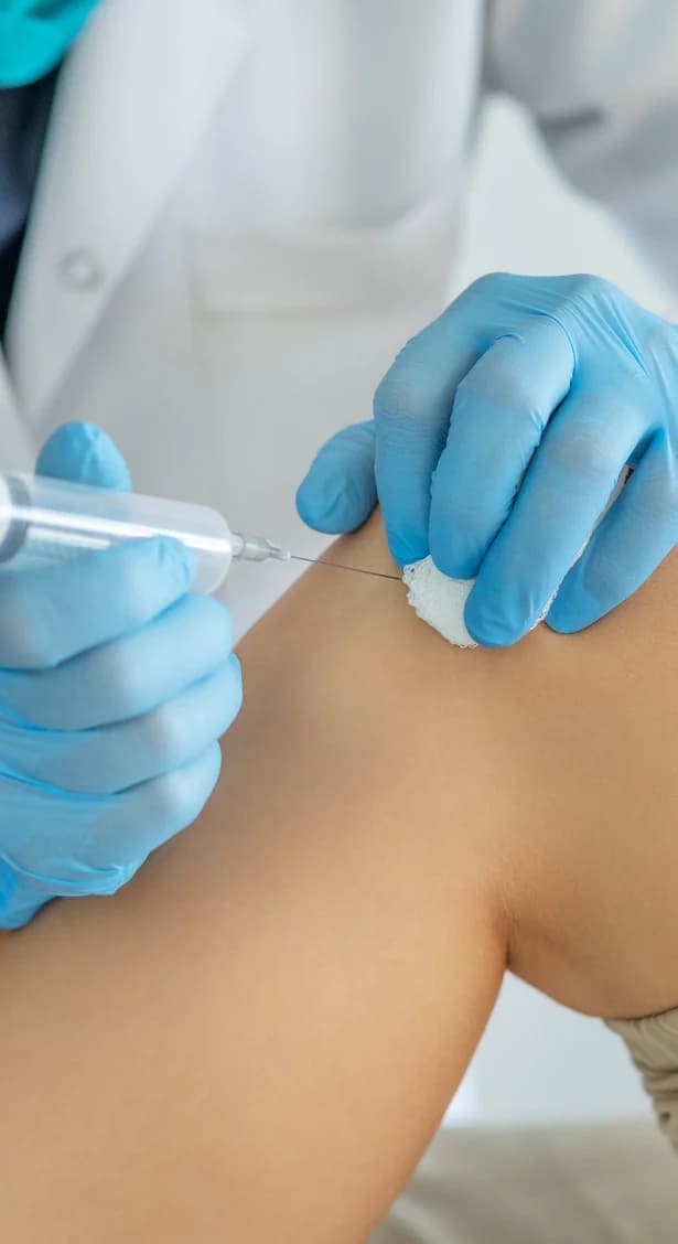
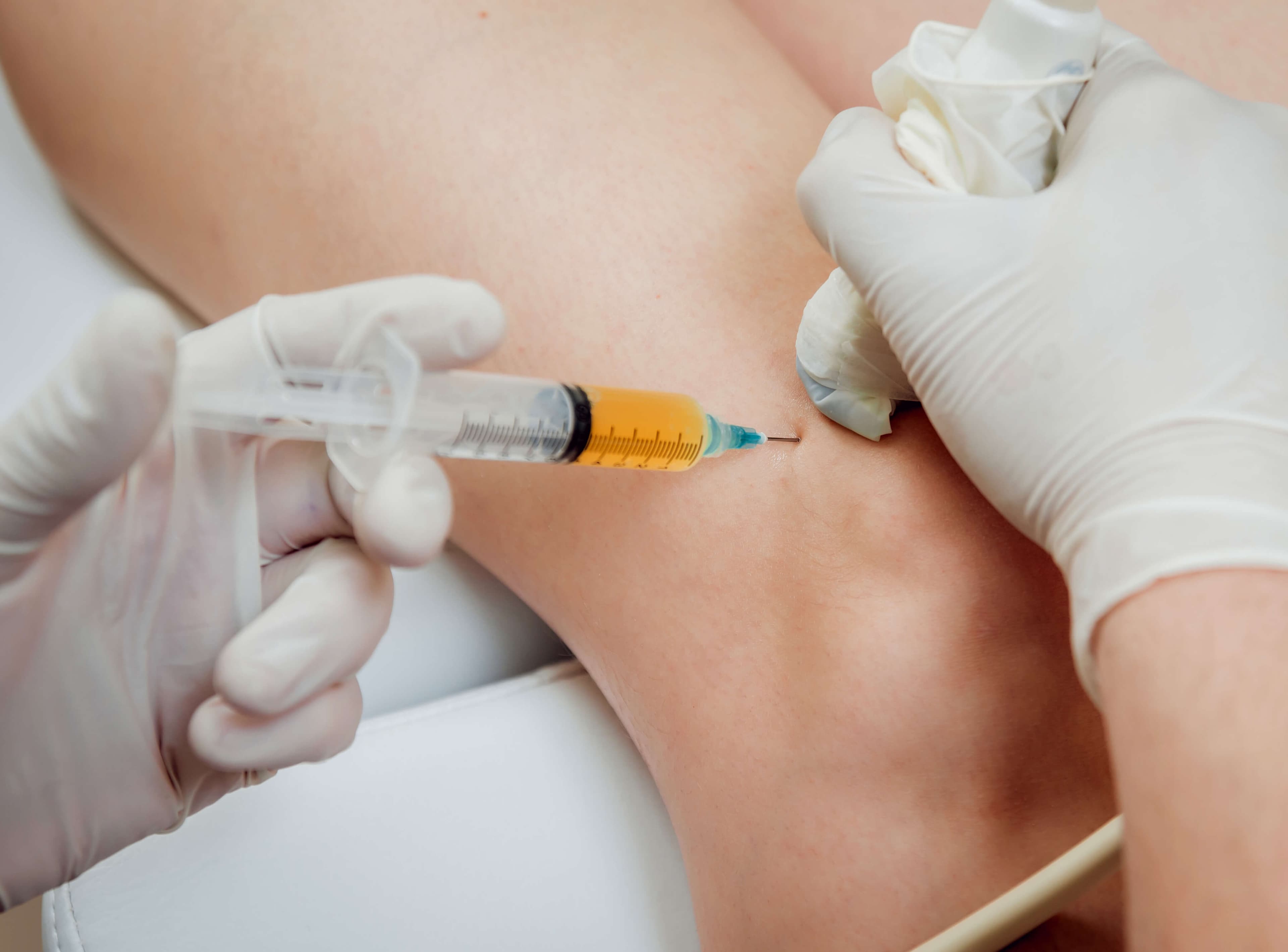
Clinician delivering a prepared joint injection
Delivery to the target joint in the same sitting.

Tissue-specific

Auricular cartilage supplies the autologous starting material.

Mechanical only

No culture, enzyme digestion or ex-vivo expansion.

Point of care

Sampling, preparation and delivery happen in one clinical pathway.

The NanoACi sequence

Three biological roles. One defined clinical sequence.

NanoACi is more than a list of components. Patient selection, preparation order, timing, image-guided delivery and aftercare make it a reproducible surgeon-led technique.

01

Cellular step

Autologous micrografts

Tiny samples of the patient’s auricular cartilage are mechanically prepared at point of care using the Mytocel MSK process. Nothing is laboratory-cultured over several weeks.

02

Scaffold step

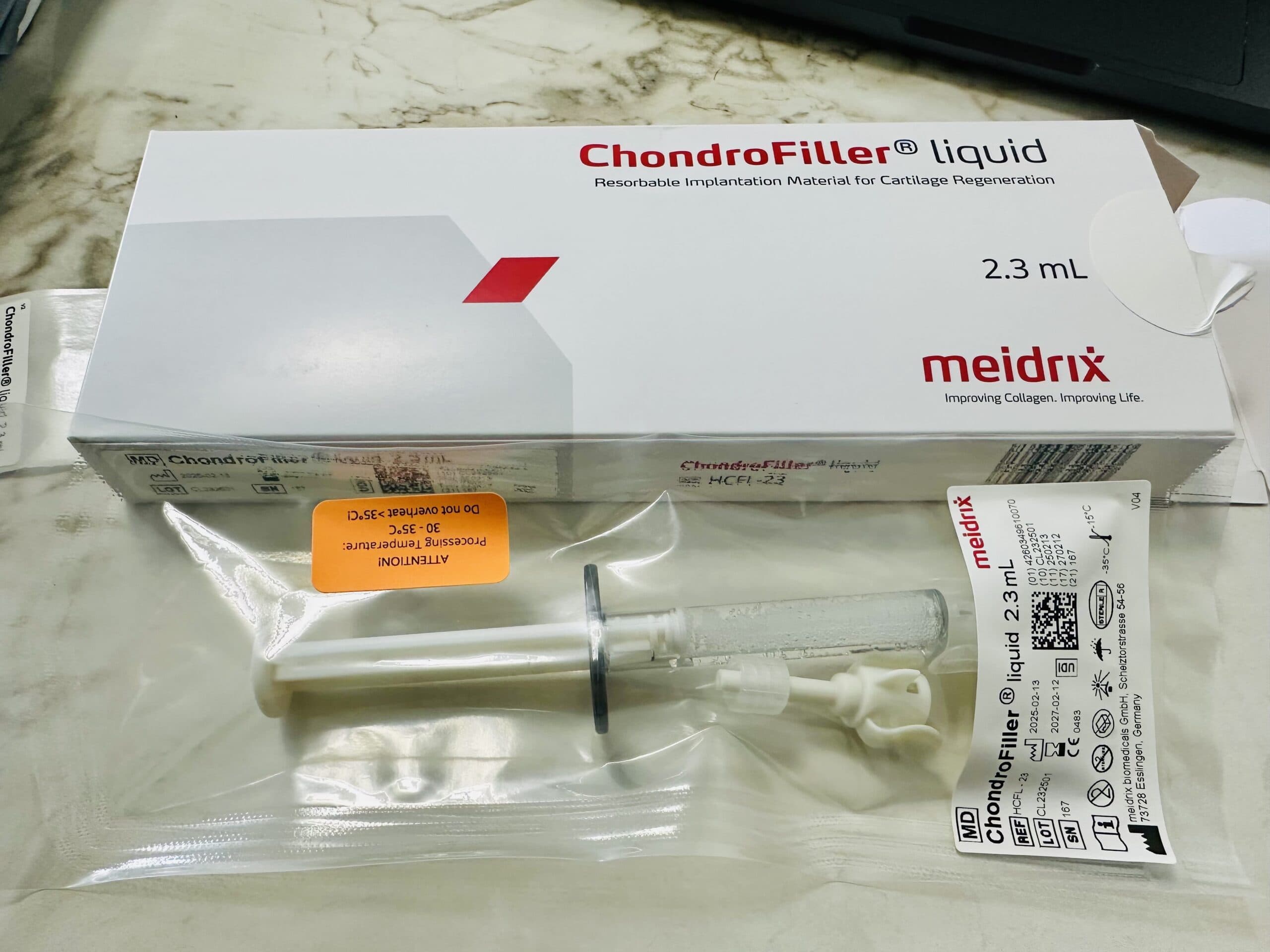
ChondroFiller

A cell-free Type I collagen hydrogel scaffold manufactured by Meidrix Biomedicals GmbH provides the structural matrix.

03

Biological-support step

Platelet-rich fibrin (PRF)

PRF is prepared from the patient’s own blood and supplies blood-derived biological support within the sequence.

What happens in practice

From scan review to needle delivery.

  1. 01

    Define the joint problem

    Examination and imaging establish the cartilage pattern, joint mechanics and whether an injectable preservation pathway is a reasonable option.

  2. 02

    Prepare the cellular step

    Auricular cartilage is prepared into autologous micrografts at point of care using the Mytocel MSK process.

  3. 03

    Prepare scaffold and PRF

    The cell-free ChondroFiller collagen scaffold and platelet-rich fibrin from the patient’s blood keep separate, defined biological roles.

  4. 04

    Deliver the defined sequence

    Professor Lee delivers the planned components by image-guided needle technique in one sitting, without arthroscopy.

The diagram is an orientation, not a substitute for Professor Lee's individual procedural plan. Exact preparation and delivery details remain part of the clinical protocol.

Four-step NanoACi guide showing preparation, auricular cartilage sampling, point-of-care preparation and joint delivery
NanoACi keeps cartilage expertise and specialist judgement while changing the route of delivery: one stage, needle-delivered and non-arthroscopic.
Evidence maturity

What the evidence supports, layer by layer

Component studies and complete-protocol outcomes answer different questions. They are kept separate here.

01

Published component evidence

Mytocel MSK component

Published studies describe the autologous micrograft technology across musculoskeletal applications. Those studies support the cellular component; they are not studies of the complete NanoACi sequence.

Read the source evidence

02

Published device evidence

ChondroFiller component

ChondroFiller has a device-specific evidence base across cartilage applications. This evidence relates to the scaffold and its clinical use, not automatically to every protocol in which it appears.

Read the source evidence

03

Newer combined protocol

Complete NanoACi protocol

The exact three-part NanoACi technique is newer. NanoACi 100 is the prospective programme designed to follow the first 100 consecutive cases using defined pain, function and imaging measures.

Read the source evidence

Clinical decision

Treatment names do not decide suitability.

Options that may be discussed include NanoACi, ChondroFiller, Mytocel MSK, established conservative care, surgery or joint replacement, depending on examination and imaging.

The relevant questions include the diagnosis, pattern and depth of cartilage damage, joint stability and alignment, symptoms, previous treatment and the full set of realistic alternatives.

FAQ

NanoACi and Mytocel MSK questions

Stable definitions and evidence boundaries for the most common questions.

NanoACi is Professor Lee’s single-session injection technique. It sequences autologous cartilage micrografts, PRF prepared from the patient’s blood and a ChondroFiller collagen scaffold. NanoACi describes the technique, not a manufactured product.

Mytocel MSK is an autologous micrograft treatment. Tiny samples of the patient’s auricular cartilage are mechanically prepared at point of care into micrografts without enzyme digestion or laboratory cell culture. It can be considered independently and is also the cellular step within NanoACi.

No. Mytocel MSK supplies autologous micrografts. NanoACi is the wider Professor Lee technique in which that cellular step is delivered with PRF and a ChondroFiller scaffold in a defined single-session sequence.

Mytocel MSK uses tiny samples of auricular cartilage as accessible, tissue-specific starting material. The samples are taken under local anaesthetic and mechanically prepared at point of care; cartilage is not harvested from the damaged joint for this step.

No. Mytocel MSK is an autologous micrograft treatment. The auricular-cartilage samples are mechanically disaggregated without enzyme digestion, laboratory culture or ex-vivo expansion, then delivered within the same clinical pathway.

After assessment and sterile preparation, the auricular-cartilage micrograft component and PRF from the patient’s blood are prepared at point of care. ChondroFiller provides the separate cell-free collagen scaffold. Professor Lee then delivers the defined protocol by image-guided needle technique without arthroscopy.

ChondroFiller is the cell-free collagen scaffold within NanoACi. It can also be delivered independently as an ultrasound-guided injection or used surgically within a cartilage-repair pathway such as Liquid Cartilage.

The autologous micrograft, PRF and ChondroFiller components each have relevant evidence. The exact combined NanoACi protocol is newer, so component evidence and complete-protocol evidence are reported separately. NanoACi 100 is designed to collect prospective outcomes for the full technique.

Suitability cannot be decided from a website or treatment name. Professor Lee considers the diagnosis, examination, imaging, distribution of cartilage damage, joint mechanics, overall health and realistic alternatives before discussing any injection, operation or replacement pathway.

The treatment follows the diagnosis

Imaging, examination and realistic alternatives come first

NanoACiMytocel MSKChondroFillerCartilage preservation

Options that may be discussed include NanoACi, ChondroFiller, Mytocel MSK, established conservative care, surgery or joint replacement, depending on examination and imaging.

Privacy & Cookies Policy